One concern we have in veterinary science is the lack of understanding in our profession when it comes to liver cancer, and the association pet owners may have with humans when it comes to this disease. Liver cancer is surprisingly curable in many cases when it comes to our cats and dogs.
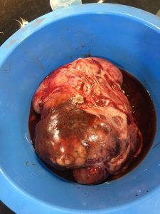
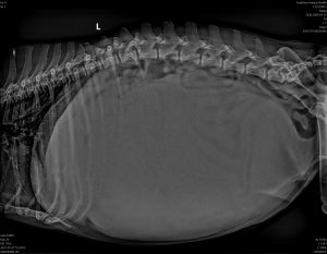
Max’s case highlights both the potential nonunderstanding of this condition by both veterinarians and pet owners alike. Max, a 14 year old Maltese cross, came to us for a second opinion having been diagnosed with an inoperable liver cancer, and had been recommended euthanasia. You can see from both the X-Ray, and the mass removed post surgery (GRAPHIC IMAGE WARNING, scroll down to view), that this was a large cancer, taking up most of the room in Max’s abdomen. It was no wonder Max was inappetant and lethargic. However the basics of a liver mass, when first presented with one, are the following:
- Until we have a sample we don’t know for sure what it is i.e. cancer, abscess or an inflammatory lesion. (Even though we can guesstimate it likely a cancer of some sort).
- If it is a cancer we don’t know if it’s already spread, or likely to return even if removed, as that’s dependent on what type of cancer it is, and how advanced it is. What we do know is, the sooner we get rid of it the less likely it is to spread.
- Many liver masses (even highly likely cancerous ones) are potentially removable, and depending on what type of cancer, it is either significantly palliative (i.e.: the patient will live a happy and great life for some years before it returns), or even curable.
So what can we do when presented with a case like Max’s? We recommend the following:
1) Stage the dog: This means look to see if we can see signs that if it is a cancer it has spread, and check overall bloods to ensure no other significant problems. This can be done by full body CT scan ($2,200 at Southern Animal Health), or advanced radiology and ultrasound ($880 at Southern Animal Health).
2) If staging is clear, we recommend an exploratory laparotomy to assess the nature of the mass, the rest of the abdomen, and the potential to surgically remove the mass.
3) Before doing this surgery we have a long discussion with owners about what to potentially expect using two of the most common cancers at either end of the spectrum as examples. As such we discuss some of the following points:
- Haemangiosarcomas are a common cancer that have a certain look and feel about them that would make us highly suspicious upon direct examination. Unfortunately these are difficult to remove from the liver with about a 50% success rate only, and most will return within a few months to possibly one year even if surgery is successful. But it’s still possible to get a result. (Very different to splenic haemangiosarcomas, or haemangiomas, which carry a 95% excellent prognosis to get through surgery with no problems at all, but also with a much better potential cure rate than hepatic ones).
- Hepatic adenocarcimoma is nasty as a local cancer, but is far easier to remove with a 95% success rate if in the left lobes ( a little less if in the right lobes). These cancers also carry about a 50% chance of cure, or if not cured by surgery may be years before they return , with an otherwise happy and healthy dog until that time. Without removal they are painful and cause the demise of the dog.
4) So with these two possible extreme examples explained, owners are well informed to make an in-surgery decision about what they might do once we are having a look. This is a very important point about our exploratory laparotomies at Southern Animal Health: We have owners involved during the surgery to inform them exactly what we have, and what the prognosis is. That allows them to be part of a sensible decision, having been well prepared before surgery with various scenarios we may encounter.

Max’s 2kg Liver Mass
With this in mind Max’s owner, not having had any of this given as an option, wanted to try anything possible once we had this lengthy discussion, and wanted to be part of the decision process once we knew exactly what was going on. Grading proved clear, exploratory laparotomy revealed a single lobe resectable mass with the rest of the abdomen clear. We removed a 2kg mass from a dog weighing 8kg pre surgery and then 6kg’s post surgery. Proportionally to the size of the dog, this was one of the largest liver cancers we have removed.
Max recovered really well from surgery, eating like he hadn’t for a long time the very next day. Although we were hopeful we may have been able to resect the whole mass, we never know until pathology comes back. Max’s pathology came back as a hepatic adenocarcinoma, with a clear margin! The prognosis for cure here is high. Two months post surgery Max is living life to the full, with hopefully years to come despite being 14 years at the time of diagnosis. If you have a pet diagnosed with possible liver cancer, no matter how large or aggressive it is, please understand you have options.

Max’s liver cancer – taking up most of the room in his abdomen
Note for Veterinarians:
Chronic Active Hepatitis: If you are ever doing an exploratory surgery to examine and biopsy a liver for liver disease, CAH is an autoimmune inflammatory disease that often makes the liver look like it has serious cancer throughout the whole liver. We have heard of many of these cases being euthanised on the table due to the suspicion of it looking like it has cancer right through the liver. These often present with hundreds of medium nodules with no normal looking liver left. Please biopsy these, as most times it is not cancer, it’s CAH, and can be managed or even cured with steroids. Do not recommend euthanasia on the table.
